The Eye
Cones and Rods and Retina
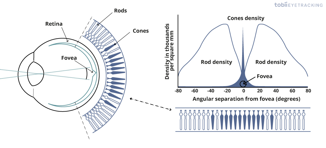
Cones and rods are the light sensitive cells which combine to form the retina at the back of the eye, and form the basis for the brain’s ability to measure and thus perceive light and images.
Cones are tuned for bright lights, color measurements, and high amounts of visual detail. Cones are more concentrated at the center of our visual perception, allowing us to read and do other highly detail-oriented tasks.
Rods are tuned for low-light conditions, cannot see color, and are more tuned for movement-awareness than high detail. Rods are more concentrated in your periphery vision, which is why your vision in the dark is actually more detailed along the sides of your vision than straight ahead.

The Fovea
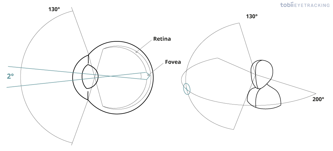
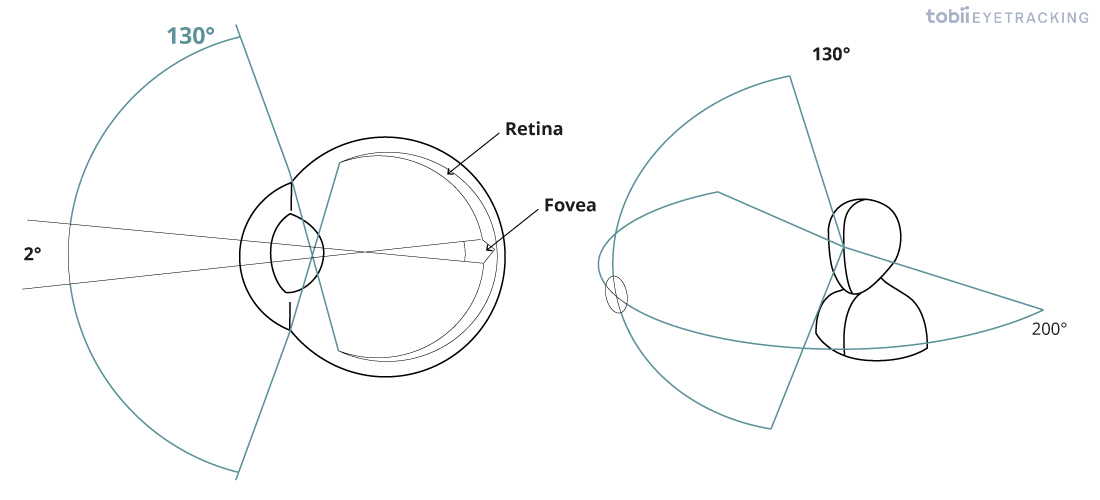
There is a small part of the retina called the fovea, where we have the highest concentration of cones, leading to the highest visual acuity and color perception. The fovea is in the center of our vision and can only perceive a small region, about 2°, which is roughly the size of the thumbnail when the arm is extended fully forward.
It is here where most of our conscious vision occurs, and this is the only area in which we have the ability to read text. You can test this yourself by staring at any word and seeing how many words you are able to read outside of the center of your eye. You won’t be able to read words much outside of a visual angle of 2.5°, and it takes 25% of the brain’s visual processing power to turn these 2.5° into perceivable text, which is remarkable given our 200° by 130° field of view.

Peripheral Vision
Our peripheral vision is still highly sensitive to motion, flickering, contrast changes and sharp edges, not all of which we will be conscious of, but all are capable of triggering rapid sub-conscious re-orientation of the eyes or ‘saccades’. Moving imagery which generates too many saccades can be uncomfortable to watch and can feel like having dust in your eye.
Our sense of having a complete detailed view of the world is maintained by our brain using these limited inputs, small rapid involuntary eye movements (micro-saccades), full saccades, memory and sensitivity to motion and contrast changes.

Focus with Cornea and Lens Accommodation
When light enters the eye, both the cornea and lens focus light onto the retina, which is the light-sensitive tissue at the back of the eye that enables the brain to perceive images. The cornea does roughly two-thirds of all the focusing and is unable to change shape.
Irregularly shaped corneas can be the cause of many eye-sight-related issues such as nearsightedness, farsightedness, and astigmatism. The remaining one-third of the focus is done dynamically with our lens, which has the ability to be stretched by little muscles in our eye to create crisp images at many distances. This action of focusing with our lens is called lens accommodation.
